Seborrheic keratoses appear “stuck-on” with uniform colour, while melanomas look integrated into the skin with multiple colours and irregular borders. See a doctor for any rapidly changing or unusual growths.
- Dr Sharon Crichlow
- Reading Time: 10 Mins
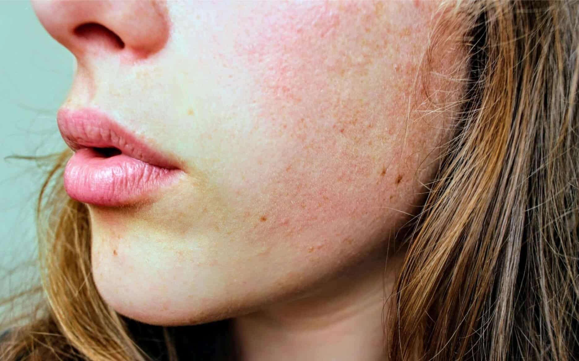
Spotting the difference between a harmless skin growth and something more serious can be challenging. Seborrheic keratosis and melanoma can appear similar to untrained eyes, but knowing the key differences could literally save your life. This guide will help you understand what to look for and when to seek professional advice.
Key Takeaways
- Seborrheic keratoses are benign growths with a characteristic “stuck-on” appearance, while melanomas grow into the skin with irregular features.
- Use the ABCDE rule (Asymmetry, Border, Colour, Diameter, Evolution) to identify potential melanomas requiring medical attention.
- When in doubt, always seek professional evaluation; 0.66% of lesions diagnosed as seborrheic keratosis were actually melanoma in clinical studies.
Table of Contents
What Is Seborrheic Keratosis?
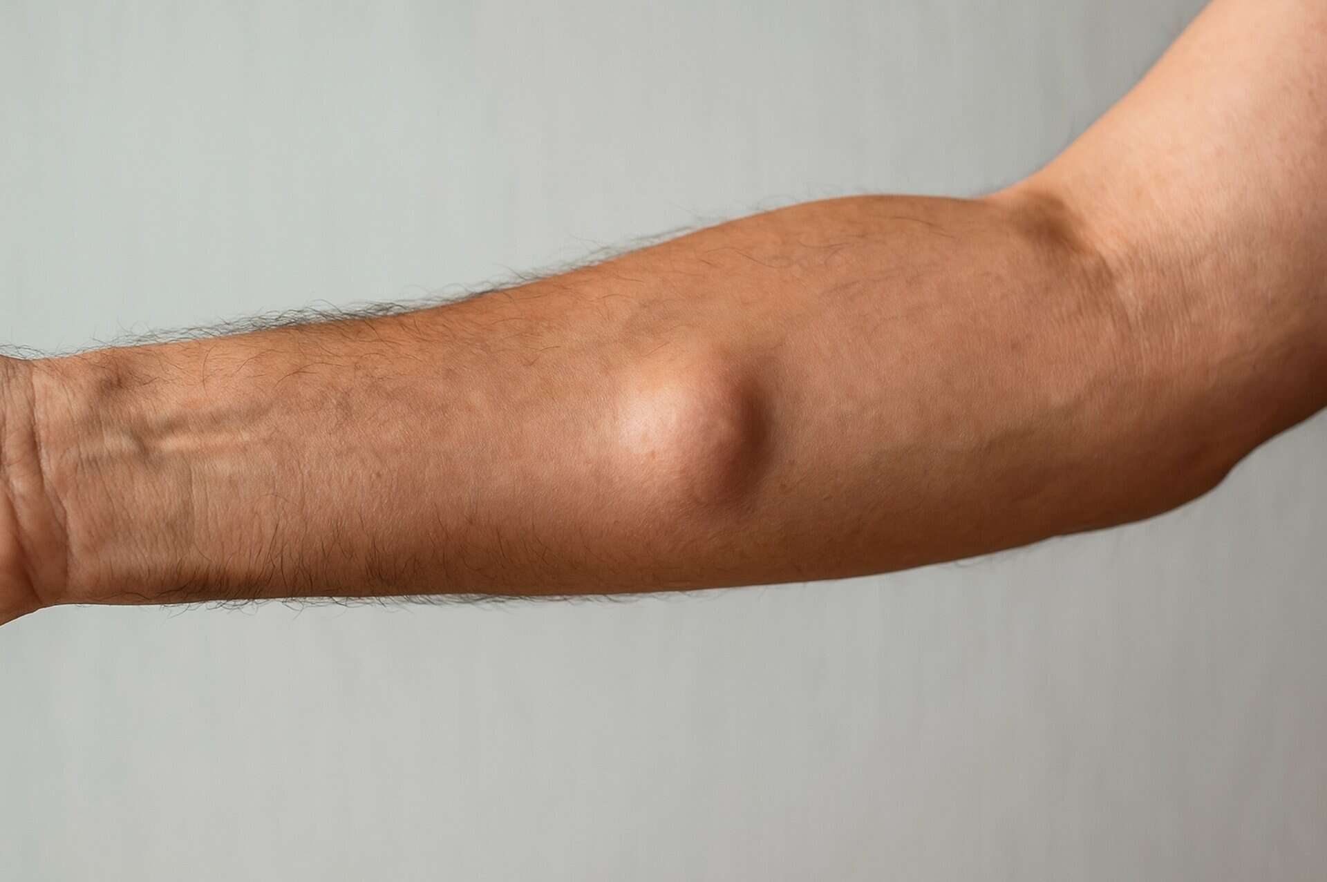
Seborrheic keratosis (SK) is a common, completely benign skin growth that typically appears during middle age or later. These growths develop when skin cells called keratinocytes multiply excessively, creating raised lesions on the skin’s surface.
They often look like they’re “stuck on” to your skin, as if you could flick them off with your fingernail. Despite sometimes appearing concerning due to their dark colour or rough texture, seborrheic keratoses are not cancerous and cannot become cancerous.
Most people will develop multiple seborrheic keratoses throughout their lifetime, with their numbers increasing with age. By age 70, more than 80% of individuals will have at least one seborrheic keratosis.
What Is Melanoma?
Melanoma is the most dangerous form of skin cancer. It develops when melanocytes, the cells responsible for skin pigmentation, grow uncontrollably and form malignant tumours.
Unlike seborrheic keratosis, melanoma can spread (metastasise) to other parts of the body if not detected and treated early. This aggressive spread makes early identification crucial for successful treatment.
Melanoma accounts for only about 4% of all skin cancers, but causes the majority of skin cancer deaths. The good news is that when caught early, melanoma has a very high cure rate.
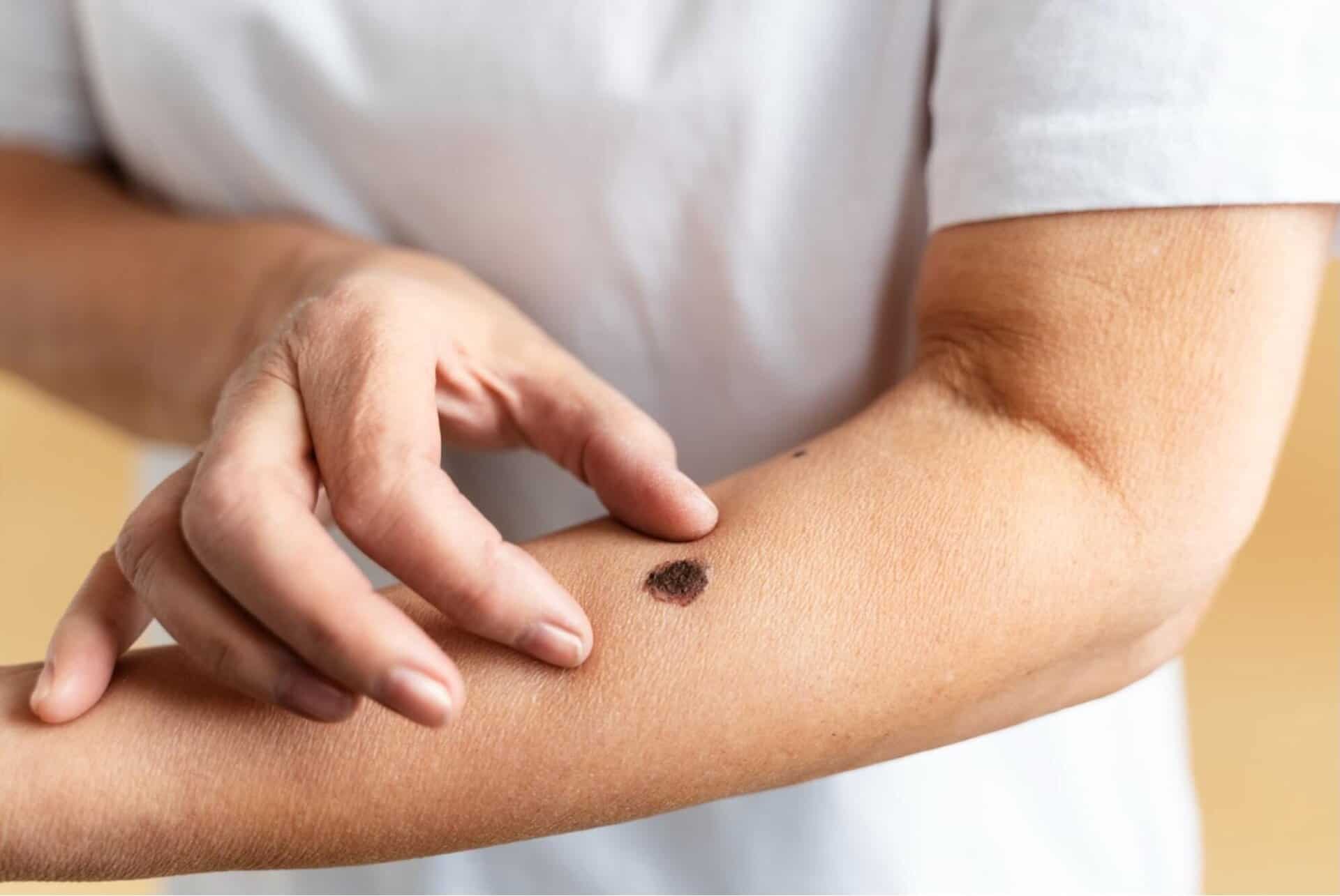
Key Visual Differences Between Seborrheic Keratosis and Melanoma
Learning to spot the differences between seborrheic keratosis and melanoma could potentially save your life. Here are the key visual distinctions to watch for:
| Feature | Seborrheic Keratosis | Melanoma |
|---|---|---|
| Appearance | "Stuck-on" appearance, as if pasted onto skin | Integrated with skin, growing into rather than on top of skin |
| Shape & Symmetry | Usually symmetrical, round or oval | Often asymmetrical with irregular shape |
| Colour | Generally uniform colour (tan to black) | Multiple colours within same lesion (brown, black, red, blue, white) |
| Borders | Well-defined, clear edges | Irregular, notched, or blurred borders |
| Texture | Rough, warty, or waxy surface | May be smooth or varied texture |
| Surface | Often has visible grooves or ridges | May be flat or have irregular surface |
| Growth Pattern | Slow, gradual growth | May change rapidly in size, shape, or colour |
Healthcare professionals often use the ABCDE rule to identify potential melanomas:
- Asymmetry: Uneven shape
- Borders: Irregular or blurred edges
- Colour: Multiple colours or uneven distribution
- Diameter: Larger than 6mm (pencil eraser)
- Evolution: Changes in size, shape, colour, or symptoms
If you’re concerned about any suspicious moles or skin growths, our comprehensive mole checking service provides expert evaluation with same-day results in many cases.
Common Locations and Appearance Variations
Seborrheic keratoses most commonly appear on the trunk (chest and back), face, neck, and shoulders. They rarely develop on the palms, soles, or mucous membranes.
Their appearance can vary widely. Some are flat while others are raised; colours range from light tan to dark brown or black. Some have a smooth surface while others appear rough and warty.
Melanomas can develop anywhere on the body, including areas not regularly exposed to the sun. Common locations include the back, legs, arms, and face. In men, the trunk is a common site, while in women, the legs are frequently affected.
Melanomas in people with darker skin tones often appear on the palms, soles, and under nails, areas with less pigmentation.
Risk Factors for Melanoma
Several factors can increase your risk of developing melanoma. UV exposure is perhaps the most significant risk factor. A history of sunburns, especially during childhood, and regular use of tanning beds significantly increases melanoma risk.
Individuals with fair skin, light-coloured eyes, and red or blonde hair have less protective melanin and are at higher risk. However, people with darker skin tones can also develop melanoma.
Having numerous moles (more than 50), especially abnormal ones, increases risk. Those with a personal or family history of melanoma or other skin cancers should be particularly vigilant.
People with weakened immune systems due to conditions like HIV/AIDS, certain medications, or organ transplants face elevated risk. Age also plays a role, with risk increasing after age 50, though melanoma also affects younger adults.
Causes and Risk Factors for Seborrheic Keratosis
Unlike melanoma, seborrheic keratosis is not caused by sun exposure, though the exact cause remains unclear. Age is the primary risk factor, with prevalence dramatically increasing after age 50. By age 70, more than 80% of individuals will have at least one seborrheic keratosis.
Genetic factors appear to play a role, as SK often runs in families. Some research suggests hormonal influences may contribute, as these growths sometimes appear or increase during pregnancy.
A common misconception is that seborrheic keratoses can become cancerous. Research consistently shows they have no malignant potential whatsoever.
When to See a Doctor
Seeking timely medical evaluation is essential when you notice concerning skin changes.
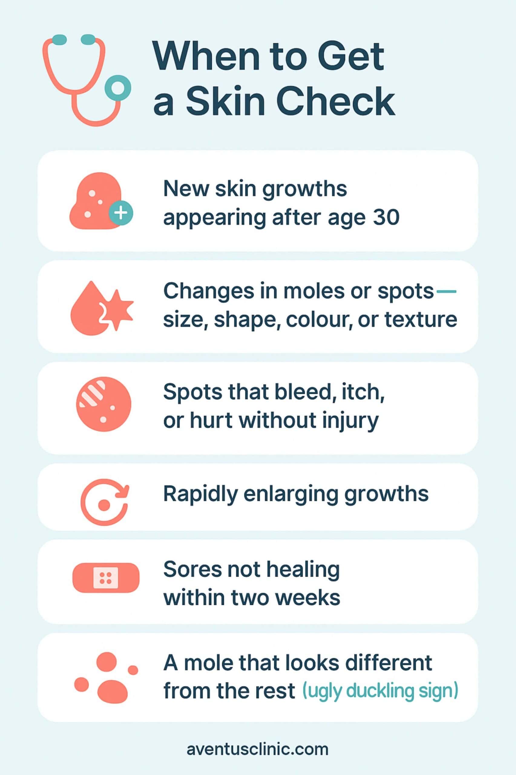
According to a JAMA Dermatology study, 0.66% of specimens clinically diagnosed as seborrheic keratosis were actually melanoma upon pathological examination. This underscores the importance of professional evaluation.
If you’re concerned about any skin changes, our experienced dermatologists in Hertfordshire can provide a comprehensive assessment using advanced diagnostic techniques.
Treatment Options for Seborrheic Keratosis and Melanoma
Treatment approaches differ dramatically between these conditions due to their fundamental difference in nature.
- Seborrheic Keratosis Treatment: Since seborrheic keratoses are benign, treatment is optional and often performed for cosmetic reasons or if lesions become irritated. Common removal methods include cryotherapy (freezing), curettage (scraping), electrosurgery, shave excision, or laser therapy.
- Melanoma Treatment: Melanoma treatment depends on the stage but almost always involves surgical removal of the cancer and surrounding tissue. Early-stage melanomas may require only surgery, while advanced cases might need additional treatments like immunotherapy, targeted therapy, radiation, or chemotherapy.
For safe and effective mole removal in Hertfordshire, we offer both cosmetic and medical removal options performed by qualified specialists using the latest techniques to minimise scarring and ensure comprehensive histological analysis when needed.
When Seborrheic Keratosis Might Be Confused with Melanoma
According to a 2021 study published in Cancers (MDPI), approximately 40% of melanomas that resemble seborrheic keratosis are initially misdiagnosed as benign. This occurs because some melanomas can display SK-like features.
Particular caution is warranted for:
- Solitary lesions with SK features in sun-protected areas
- SK-like growths in younger individuals (under 40)
- Lesions showing recent changes in appearance
- Growths with mixed characteristics of both SK and melanoma
Advanced diagnostic technologies like reflectance confocal microscopy (RCM) and optical coherence tomography (OCT) have improved the ability to distinguish between these conditions without invasive procedures.
Conclusion
When it comes to skin lesions, knowledge and vigilance are your best protection. While seborrheic keratoses are completely harmless, their similarity to melanoma makes professional evaluation essential when in doubt.
So, are you uncertain about a skin growth? Take our free online assessment to receive expert guidance on your specific situation and determine whether you should schedule an in-person consultation with one of our skin specialists.
FAQs
Can seborrheic keratosis turn into melanoma?
No, seborrheic keratosis cannot transform into melanoma. These are completely different types of skin growths with distinct cellular origins. Seborrheic keratoses are benign overgrowths of epidermal cells with no malignant potential.
How to tell a mole, seborrheic keratosis, or melanoma apart?
Moles are typically smooth, symmetrical, and uniform in colour. Seborrheic keratoses have a “stuck-on” appearance with waxy, scaly texture. Melanomas often show ABCDE warning signs: asymmetry, irregular borders, colour variation, diameter >6mm, and evolution over time.
Are seborrheic keratoses hereditary?
There appears to be a genetic component to seborrheic keratosis development, as they tend to run in families. However, no specific genetic markers have been definitively identified, and environmental factors likely play a role as well.
Can melanoma appear in areas not exposed to the sun?
Yes, melanoma can develop anywhere on the body, including areas rarely exposed to sunlight, such as the soles of the feet, palms, under nails, or between toes. About 20-30% of melanomas develop in sun-protected areas.
What should I do if I notice a suspicious lesion?
If you notice any concerning skin changes, consult a dermatologist promptly. Don’t try to self-diagnose or remove suspicious lesions. Professional evaluation using dermoscopy and other advanced diagnostic techniques provides the most accurate assessment.